lead i ii iii placement, check these out | Where are leads I II and III placed?
Where are leads I II and III placed?
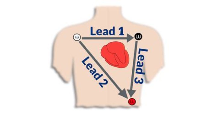
Leads I, II, III, aVF, aVL and aVR are all derived using three electrodes, which are placed on the right arm, the left arm and the left leg. Given the electrode placements, in relation to the heart, these leads primarily detect electrical activity in the frontal plane.
What are leads I II and III?
In the lead II configuration, the positive electrode is on the left leg and the negative electrode is on the right arm. Lead III has the positive electrode on the left leg and the negative electrode on the left arm.
What is the correct lead placement?
Precordial Lead Placement
V1 is placed to the right of the sternal border, and V2 is placed at the left of the sternal border. Next, V4 should be placed before V3. V4 should be placed in the fifth intercostal space in the midclavicular line (as if drawing a line downwards from the centre of the patient’s clavicle).
Where is the V3 lead placed?
V3 sits midway between V2 and V4. Follow the 5th intercostal space to the left until your fingers are immediately below the beginning of the axilla, or under-arm area.
Where should cardiac monitoring leads be placed?
Place the left arm (LA) electrode near the left shoulder, close to the junction of the left arm and torso. Place the right leg (RL) electrode below the level of the lowest rib on the right abdominal area. Place the left leg (LL) electrode below the level of the lowest rib on the left abdominal area.
Where do female ECG leads go?
Electrode placement for women
Explain to the patient what you plan to do in terms of electrode placement; emphasize that several of the chest leads may need to be placed around and under the left breast.
Which ECG machine is best?
Healthline’s picks for the best ECG devices
EMAY Portable ECG Monitor.1byone Portable Wireless ECG/EKG Monitor.Omron Complete Wireless Upper Arm Blood Pressure Monitor + EKG.Eko DUO ECG + Digital Stethoscope.Biocare 12-Lead ECG Machine.Omron KardiaMobile EKG.Wellue Portable EKG Monitor.
Why do we use lead II for ECG monitoring?
To assess the cardiac rhythm accurately, a prolonged recording from one lead is used to provide a rhythm strip. Lead II, which usually gives a good view of the P wave, is most commonly used to record the rhythm strip.
What is a 3 lead ECG used for?
3-lead ECGs are used most often for recording a 24-hour reading. A 24-hour reading is a frequently used tool for the diagnosis of heart problems and is reimbursed as a long-term reading.
How do you place a 3 lead ECG?
Position the 3 leads on your patient’s chest as follows, taking care to avoid areas where muscle movement could interfere with transmission:
WHITE.RA (right arm), just below the right clavicle.BLACK.LA (left arm), just below the left clavicle.RED.LL (left leg), on the lower chest, just above and left of the umbilicus.
When is a 15 lead ECG used?
Therefore, the use of the 15-lead ECG may confirm the STEMI diagnosis while determining its actual extent. The term “posterior infarction” identifies an AMI that insults the left ventricular wall by occlusion of the right coronary artery–posterior descending branch or the circumflex artery (15).
When is an 18 lead ECG used?
18-lead synthesized ECG is expected to be useful in detecting right side and posterior infarction. Guidelines such as AHA, ACC or ESC recommend to measure additional lead (V3R-V5R and V7-V9) for the patient with suspected acute coronary syndrome.
How do you know if ECG leads are incorrectly placed?
Lead reversals do happen; the most common is right and left arm reversals. Your first clue is a negative QRS complex in lead I. A predominantly upward P-QRS-T complex in aVR is another big clue. When in doubt, repeat the ECG!
Where are the 12 leads placed on a patient for an ECG?
Electrode placement for a 12-lead ECG is standard, with leads placed on the left and right arm and left and right leg. Another pair of electrodes is placed between the fourth and fifth ribs on the left and right side of the sternum.
Where do electrodes go on ECG?
Simple steps for the correct placement of electrodes for a 12 lead ECG/EKG:
Prepare the skin. Find and mark the placements for the electrodes:First, identify V1 and V2. Next, find and mark V3 – V6. Apply electrodes to the chest at V1 – V6. Connect wires from V1 to V6 to the recording device. Apply limb leads.
What are the two leads we should be monitoring most patients in?
Multiple lead monitoring is superior to single lead monitoring. If two leads are available, V1 and lead III or aVF (or a limb lead with maximal ST segment displacement) are good choices. If three leads are available, leads V1, III, and aVF are the best choices.
Related Archive

harry potter trivia show host, latest free online harry potter movies, best HD videos you should watch in 2022 – 2023

harry potter uniform pattern, latest free online harry potter movies, best HD videos you should watch in 2022 – 2023

harry potter vans ebay, latest free online harry potter movies, best HD videos you should watch in 2022 – 2023