middle scalene origin and insertion, check these out | Which is the insertion of the middle scalene?
The middle scalene arises from the posterior tubercles of the transverse processes of the lower six cervical vertebrae. It descends along the side of the vertebral column to insert by a broad attachment into the upper surface of the first rib, posterior to the subclavian groove.
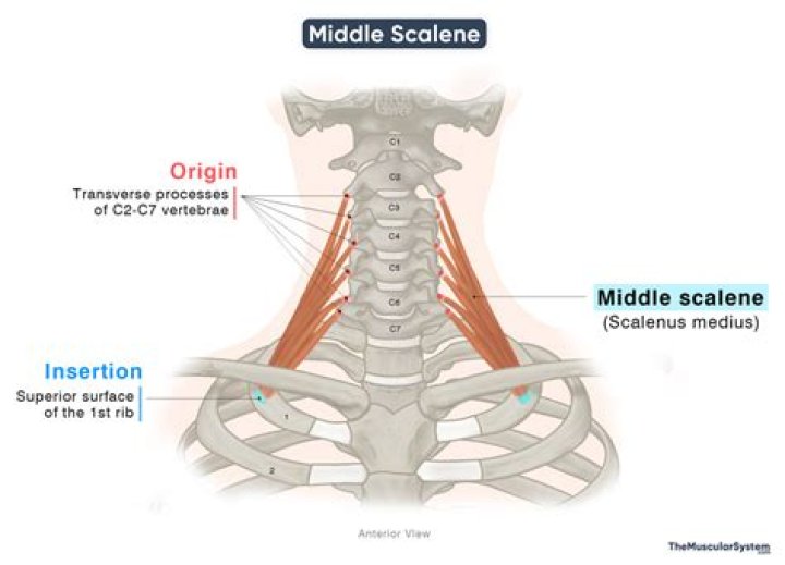
Which is the insertion of the middle scalene?
The middle scalene originates from the transverse processes of the last six cervical vertebrae, between the anterior and posterior tubercle, inserting itself on the upper face of the first rib, posterior to the sulcus of the subclavian artery.
Where do the scalenes insert?
Origin and insertion
The scalenus posterior is the smallest of the scalene muscles. Its fibers originate from the posterior tubercles of transverse processes of cervical vertebrae C5-C7. The muscle extends posterolaterally and tapers into a thin tendon, which inserts into the external surface of the 2nd rib.
Which is the action of the middle scalene?
Function. The action of the anterior and middle scalene muscles is to elevate the first rib, they also flexes and laterally bends the neck to same side. The action of the posterior scalene is to elevate the second rib and tilt the neck to the same side.
What pierces the middle scalene?
The middle scalene is the longest and largest of the scalene muscles. The first two roots of the long thoracic nerve and the dorsal scapular nerve pierce the middle scalene and emerge on its lateral surface.
Which is the insertion of the anterior scalene?
By a narrow, flat tendon attaches into the scalene tubercle on the inner border of the first rib, and into the ridge on the upper surface of the rib in front of the subclavian groove.
What attaches to the scalene tubercle?
First rib. The scalene tubercle is a small projection that runs along the medial border of the first rib between two grooves, which travel anteriorly for the subclavian artery and posteriorly for the subclavian vein. It projects outward medially, and is the site of insertion for scalenus anterior.
Does the middle scalene pass through the scalene gap?
The scalene triangle is bound by the anterior and middle scalene muscles, with the first rib at the base. The brachial plexus nerves (yellow) and the subclavian artery (red) pass through the scalene triangle, while the subclavian vein (blue) passes in front. Another muscle in this area is the middle scalene muscle.
Where is the posterior scalene?
Posterior Scalene, AKA Scalenus Posterior, is one of the lateral muscles of the neck, belonging to the Scalene group. It is deeply placed, lying behind Sternocleidomastoid.
What is the origin of Sternocleidomastoid?
Origin and insertion
The sternal head originates from the manubrium sterni, while the clavicular head from the medial third of the clavicle. The insertion is the lateral surface of the mastoid process of the temporal bone and the lateral half of the superior nuchal line of the occipital bone.
What causes tight scalenes?
Activities that can cause scalene muscle pain are whiplash; excessive coughing; sufferers of breathing conditions such as COPD, asthma, and emphysema; extended periods of head tilted; sleeping on stomach with head to one side; carrying something heavy such as a backpack or purse; pulling or lifting with the arms at
What nerve runs through middle scalene?
The long thoracic nerve (LTN) originates from the ventral rami of cervical nerve roots 5, 6, and 7. It traverses the middle scalene muscle in the neck, passes under the brachial plexus and over the first rib.
What is the posterior triangle of the neck?
Posterior triangle. The posterior triangle is a triangular area found posteriorly to the sternocleidomastoid muscle. It has three borders; anterior, posterior and inferior borders. The anterior border is the posterior margin of the sternocleidomastoid muscle.
What attaches to the first rib?
The costal cartilage of the first rib articulates with the manubrium of the sternum not at the top, but lower down at its broadest part. The first costal cartilage is short and massive. It hardly permits any movement, so the two first ribs, together with the manubrium, move up and down together as one solid arch.
What anatomical structure is located in front of the scalene anterior tubercle?
Likewise, the venous groove, mostly shallow, lies in front of the tubercle or the muscular impression and extends, generally, only upto the medial 2/3 of the surface. Normally, there is no separate groove for the lower trunk of the brachial plexus.
Related Archive

harry potter trivia show host, latest free online harry potter movies, best HD videos you should watch in 2022 – 2023

harry potter uniform pattern, latest free online harry potter movies, best HD videos you should watch in 2022 – 2023

harry potter vans ebay, latest free online harry potter movies, best HD videos you should watch in 2022 – 2023